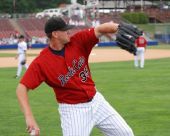
Ulnar Neuropathy In Pitchers
Jul 4, 2010 John
Conservative Management Of
Ulnar Neuropathy In Throwers
John Pallof, PT, OCS, COMT, CSCS
Introduction
Ulnar neuropathy is an increasingly common condition in throwing athletes, notably baseball pitchers. Often varying in presentation, accurate examination and diagnosis is necessary to rule out competing or concurrent elbow dysfunction. Once an accurate diagnosis of ulnar neuropathy is made, an understanding of the pathology and its mechanisms is essential to make effective treatment decisions. The goal of this article is to develop an understanding of a hypothetical model for ulnar nerve dysfunction, allowing efficient management and prevention of this condition in the the throwing athlete.
The Anatomy
The ulnar nerve typically originates in cervical nerve roots C7, C8, and T1 in varying proportions. It exits the brachial plexus as a branch off of the medial cord of the inferior trunk of the plexus. In its course away from the cervical spine it typically travels beneath, between, over, or through: the spinal foramina, the scalene complex, the clavicle, subclavius muscle, pec minor, and the latissimus tendon. The ulnar nerve then becomes firmly entrenched within the connective fascia of the neurovascular bundle, traveling below the brachial vein, anterior of the medial head of the triceps. At the insertion of the coracobrachialis it usually travels underneath the brachial vein, eventually piercing the medial intermuscular septum (approximately 8.5 centimeters proximal of the medial humeral epicondyle). From here, the nerve enters the posterior compartment of the arm, traveling through a V-shaped fascial structure composed of the medial intermuscular septum anteriorly and the Arcade of Struthers posteriorly. This structure was observed as “thin and taut,” being approximately 2 cm in length, starting 9.5 cm proximal of medial epicondyle, continuing distally to cover the ulnar nerve.

-The Brachial Plexus
At this point, an interesting change in course of the nerve takes place - it abruptly
changes direction 180 degrees to course posteriorly, creating a sort of “fold” before continuing distally. It remains in contact with the intermuscular septum, “firmly bound within connective tissue” [1]. Roughly 3 cm proximal of the medial epicondyle the ulnar nerve emerges to become superficial on its way to the retrocondylar groove in the medial elbow.
Once in the groove, the nerve is covered by a thin fascial membrane. Exiting the groove, the ulnar nerve enters the cubital tunnel between the humeral and ulnar heads of the flexor carpi ulnaris - it is covered with a “firm aponeurosis” at this point. It passes through the fibers of the FCU, emerging distally to course on top of the flexor digitorum profundis.
It is here that an important anatomical feature is present!!!! The length of the ulnar nerve between the FCU and FDP is covered by a thin fascial membrane, which is further divided into two sub-groups - one which was 5.6 cm long with three thickened fascial bands, the other being 7.7 cm long with four fascial bands.
Distally, the nerve travels through a narrow space between the pisiform and hook of hamate bones, known as Guyon’s Canal. The roof of the canal is fascial aponeurosis. The nerve finally splits into the deep and superficial branches, which are separated by a thick fascial layer.

The Concept of Neural Mobility
Oftentimes overlooked with regards to the treatment of peripheral nerve injury or dysfunction, asymptomatic nerves throughout the body have an innate amount of mobility - they need to be able to move much like the brake cable on a bike. When the brake lever is applied, the actual steel cable moves, but the plastic surrounding sheath does not. Nerves, from the spinal cord to the tiniest sensory branch, need to be able to “floss” through the various structures they travel over, under, and through. This is not hypothetical - it is well documented. The work of David Butler, McElvey, and others have generated a significant amount of knowledge and practical application in this area. Practical applications include the three primary Upper Limb Tension Tests (ULTTs) and their variants for the ulnar, radial, and median nerves; and also the Slump test positions, and their variants for sensitizing the peroneal, tibial, and sural branches peripherally. These tests are also the basis for the treatment techniques.
An interesting study specific to ulnar nerve mobility demands was performed by a team of Japanese researchers[2]. The researchers took transthoracic cadavers, and fixated their torso in an upright position - they then took the cadavers’ arms through the throwing motion, measuring the amount of movement of the ulnar nerve just proximal of the elbow, and then again at Guyon’s canal. The results are telling - an average of 12.4 millimeters at the elbow, and an average of 8.2 millimeters at the wrist. Strain within the ulnar nerve proximal of the cubital tunnel was measured to be 13.1% - “close to the elastic and circulatory limits of the nerve.” These results, combined with pre-existing knowledge of nerve pathology forms the basis for a hypothetical model of ulnar nerve dysfunction in the pitching athlete.
A Hypothetical Model
The differential in the ulnar nerve movement at the elbow and wrist suggests there is a quantifiable amount of strain that takes place in the ulnar nerve itself. Strain is defined as “deformation of a physical body under the action of applied forces”. In this case, the applied forces are the differential amounts of nerve movement taking place, and the compressive forces the ulnar nerve experiences as it rounds the elbow through the cubital tunnel. The important concept to understand is this:
If the ulnar nerve moves 12.4 mm just proximal of the elbow, and roughly 8mm at the wrist, then one can infer that a quantifiable amount of strain occurs in the ulnar nerve between these two points. This was measured to be roughly 13%!!
My theory is this: if there is normally an average 13% strain between these two points, pathological processes occur when strain percentage increases to a point where it crosses a theoretical symptomatic threshold.
The numerical value of this threshold is open to debate, but clinically it is easy - it is when the patient is experiencing symptoms. Symptoms can include (but not limited to): intermittent to constant paresthesias in ring and pinky fingers (can be especially pronounced upon wake up); pain in medial elbow - again, intermittent to constant; “pulling” sensations in any of medial elbow/upper arm, medial to ventral forearm; in extreme cases, motor weakness in ulnar distribution - especially manifesting itself as weakness/atrophy in the hand (check grip strength in the hand bilaterally, look for atrophy and difficulty with 4th and 5th digit flexion).


Muscle Wasting Due To Severe Ulnar Nerve Compromise - Ulnar Nerve Transposition Revision
Tissue Mechanics At The Elbow
An interesting study from the Journal of Shoulder and Elbow Surgery[3] provides valuable insight into how the ulnar nerve, cubital tunnel, and osseous structures interact with elbow flexion and extension. Using fresh cadaver specimens, and imaging with high resolution MRI, the researchers investigated the morphology of the ulnar nerve and the cubital tunnel in three positions: full flexion, ninety degrees of flexion, and full extension. The results provide valuable insight into the processes responsible for the gradual development of ulnar neuropathy in the pitcher.
“The ulnar nerve follows a tortuous course in full extension, becomes progressively linear with incremental elbow flexion, shifts anteriorly in the cubital tunnel, and flattens against the medial epicondyle (with progressive flexion)..”
Essentially, a portion of the ulnar nerve is compressed against a bony structure, then flossed over it repeatedly at high speeds. An appropriate analogy could be pulling a garden hose around the corner of a house - you develop a compression point where the hose hits the house.
A second, equally significant source of deformation force on the ulnar nerve comes from the copious amounts of fascial tissue at any point along the course of the nerve.
“The proximal and mid portions of the cubital tunnel also change with flexion from round to elliptical. In addition, successive increases occur in the cross sectional diameter in the medial to lateral plane. The nerve is surrounded by fat through the cubital tunnel, except adjacent to the medial epicondyle.”
From another cadaver study from the European Journal of Hand Surgery[4] - this study examines the characteristics of the fascial tissue overlying the ulnar nerve for ten centimeters distal to the midpoint of the retrocondylar groove. Twenty eight cadaver specimens were examined:
“The ulnar nerve between the flexor carpi ulnaris and flexor digitorum profundis was traced distally underneath a thin fascia. The length of fascia was measured and examined for the presence of segmental fascial thickenings, referred to as ‘bands.’ Two types of fascia were found. In Type I, three bands were identified within the fascia and the mean length of the fascia was 5.6 cm. In Type II, four bands were identified and the mean length of the fascia was 7.7 cm.”
Interesting, pertinent findings. So not only are there multiple areas of the ulnar nerve essentially being interwoven with fascial tissue, but the spatial characteristics of the cubital tunnel also change with movement. And the nerve is protected by fat everywhere except where it needs it most - where it “flattens” against the medial epicondyle with progressive flexion. These areas should be considered potential sources of abnormal deformation forces exerted on the ulnar nerve, potentially increasing strain forces within the ulnar nerve, ultimately contributing to neuropathic changes in the ulnar nerve.
Treatment Strategies
The goal of treatment/prevention is simple: minimize strain forces within the ulnar nerve. This is accomplished by taking a multi-pronged approach - address all potential sources of adverse tension on the ulnar nerve.
1. Maintain Proper Spinal Segmental Mobility - especially in the lower cervical and upper thoracic spine, as this is where the ulnar nerve originates. Stiff, mildly inflammatory segments/facet joints can eventually lead to inflammatory changes in the nerve root - which can adversely effect neural mobility. This also happens to be an area that is repeatedly heavily stressed with pitchers due to the significant amount of body on head rotation with the delivery. Indirectly, maintaining mid to lower thoracic segmental mobility is also important - mobility here allows better positioning of the head and neck, safely distributing forces, minimizing degenerative stresses.
2. Maintain Proper Upper Quarter “Suspension” - proper posture in the head/neck/shoulder girdle/thoracic spine region is an important component of treating ulnar neuropathy. Being sure to address:
a. Thoracic mobility - to be able to properly position your upper body in space so everything else works right.
b. Lat and pectoralis complex soft tissue quality and length - to have the capability to position the shoulder complex in space properly. Subscapularis soft tissue quality and length are often overlooked contributors to postural dysfunction.
c. Deep cervical flexor strength and endurance - these are the core stabilizers for the upper thoracic and cervical spines, and essential for proper shoulder function.
d. Good core stability - again, for upper body positioning in space.
e. Good pulling strength - without getting too in depth, these muscles play a significant role in upper quarter postural control, and keeping things where they should be. So the athlete must be a strong horizontal and vertical puller.
3. Soft Tissue Quality - maintaining pliable fascial tissue along the course of the ulnar nerve is an absolute must!!!!!
a. Through lat tendon, subscap, pec minor.
b. Along anteromedial bulk of the triceps.
c. Distal/medial upper arm, progressing posteriorly towards medial epicondyle. This is a very common area for symptomatic dysfunction.
d. The most important area = in, and around the flexor digitorum profundis, and the flexor carpi ulnaris!! Remember - this is where the ulnar nerve becomes “sandwiched” between the two muscles, and is covered by a variable number of fascial “slings” - significant potential areas for neural mobility impairments. These are also muscles that get a significant amount of use, as they are very important stabilizers of the elbow/wrist/hand complex in the thrower - so they are quite prone to poor soft tissue quality - increases in density, and length restrictions. Look especially in the proximal musculotendinous areas, closer to the medial epicondylar area.
e. Less often, in and around the wrist, particularly at Guyon’s canal.
4. Maintaining Adequate Neural Mobility - find a qualified rehabilitation professional to perform manual neural mobilization, and teach the athlete self mobilization exercises. This is very important as it is an essential component of a. rehabilitating the symptomatic pitcher, and b. preventing future dysfunction.
Manual Ulnar Nerve Gliding - note sequential movement, gentle
ONLY TO BE DONE BY PROPERLY TRAINED REHAB PROFESSIONALS!
Ulnar neuropathy in the throwing athlete, especially the pitcher, is often very responsive to conservative treatment as outlined above, if treated early enough. Even in the healthy pitcher, it is important to treat prophylactically to prevent dysfunction before it starts. Conservative measures, especially physical therapy are often overlooked as treatment options - oftentimes, if night splinting and injections don’t work, surgery is performed - often unnecessarily - leading to a lengthy rehabilitation without guaranteed success. I have seen several which required a revision. The above identified areas are a good place to start in rehabilitation and prevention, and should be incorporated into the pitcher’s regular maintenance program.